Philpatrick
-
Posts
68 -
Joined
-
Last visited
Content Type
Profiles
Forums
Gallery
Events
Everything posted by Philpatrick
-
Hello Jeff, The ants are a local non-invasive species. So just by chance I make observations if they happen to be around. The plant has been moved to a lower humidity environment; to see if this causes more spines to devlop than roots. The ants have gone since. Cinnamon has been used to treat orchids for fungal and bacterial problems. I use cinnamon sparingly as an ant and pest deterrent but mainly to protect flowers from being eaten by ants. Sometimes the ants eat the entire flowers of my Myrmecodia. Cinnamon oil can be phytotoxic, to some plants, so I am cautious and test it on a leaf first. Because cinnamon can be phytotoxic and can have herbicidal action, I am only going to use it as an ant barrier and not on plants. The flowering has stopped for now. A dried flower is rehydrating to get a stigma lobe count and cross section, or just wait for a new flower. I hand pollinated one flower and it looks like there is some fruit developing in a few spots. If this is fruit developing, it could be that the flowers are cleistogamous or have been pollinated by ants or an unknown pollinator. For the next flowers I am keeping the ants and possible pollinators away, and taking better notes. Are the flowers cleistogamous or do they remain closed to select a particular pollinator? Both? Protandrous? The plant is labeled "Myrmecodia cf. pulvinata".
-
I should mention that the name of this plant is noted as Myrmecodia cf. pulvinata. With a "cf." (short for, Latin: confer, "compare with").
-
Thanks Frank. I did get images of the latest flower and, hopefully, get a better understanding of the flower anatomy and breeding mechanisms. This time there was pollen and the anthers were not dehydrated and expired. Now I can see that the anthers are located at the apex of the corolla just below the petals and not low in the corolla like I thought originally, assuming all flowers are the same. The closed stigma was still developing positioned just below the anthers. The flower with one quadrant dissected to the ring of hairs. This is my view of the flower structure: It appears the unci delaminate and split leaving the upper portion open in quadrants forming the inward pointing hooks and appearance of an open flower. The lower portion of the petal delamination has an exposed cell granular surface capping off the flower; with the apex of the inner floral cavity portion remaining fused. The flowers have never opened so far on this plant. I am hoping this makes sense. Cut-away of a petal quadrant exposing a pollen sac. I have been comparing the M.tuberosa "pulvinata" drawings by Rosemary Wise to this plant, good artwork, all of it, and the flowers of this plant look different. They almost look like they have M. tuberosa "papuana" influence. With this new flower the anthers are not located in the characteristic location for M. tuberosa "pulvinata", just above the ring of hairs and are located high in the corolla. In the first flower I opened, the breeding mechanism did not seem facilitative of self fertilization, with the anthers maturing and expiring before the stigma was open and fully developed ( protandry ). I'm not sure if there is a fertilization "window" to self pollinate, but I have a creative idea to image this. In this new flower, with active anthers and pollen, the closed stigma is just below the anthers. The ant species scouting around this plant have been known to take refuge in plant cavities naturally, and is not a stinging species. I can get an image and the species name of the ants too. Perhaps not a species of ant the plant would encounter in nature but they love this plant. My guess is the debris on the flower tops is the ant refuse pile made of excavated fragments from within the alveoli cavities. I will have to look closer.


-
 A link with tuber images of Solanopteris brunei. http://herbario.up.ac.pa/Herbario/herb/vasculares/view/species/9027
 -
Here are some Microgramma bismarckii tuber images: The tuber is split vertically in line with the rhizome attachment and the entrance canal. It has been in a preservation solution. This tuber is from the same fern in the link above. The right hemisphere section in an approximate natural orientation. Right hemisphere canal detail. Left hemisphere.


-
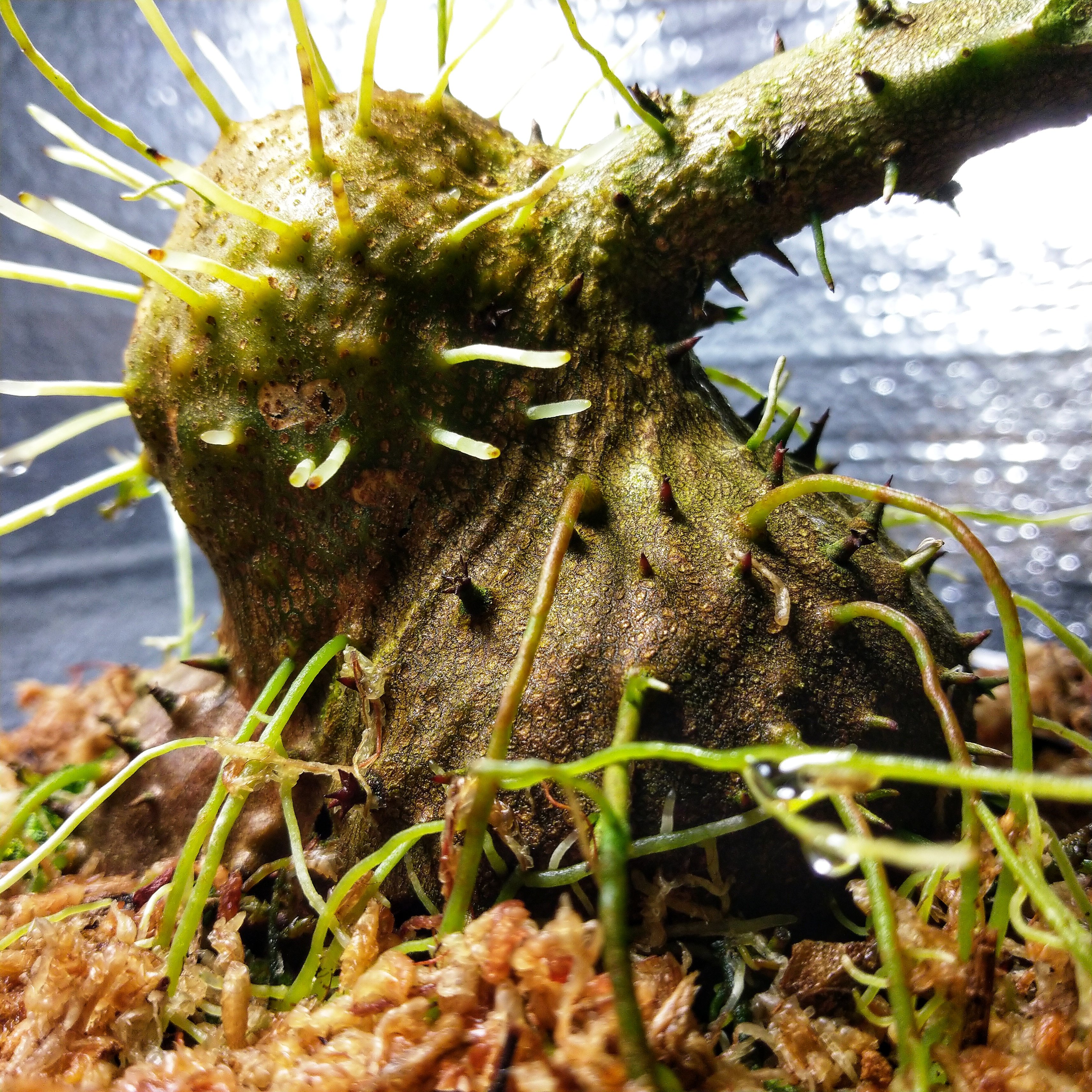
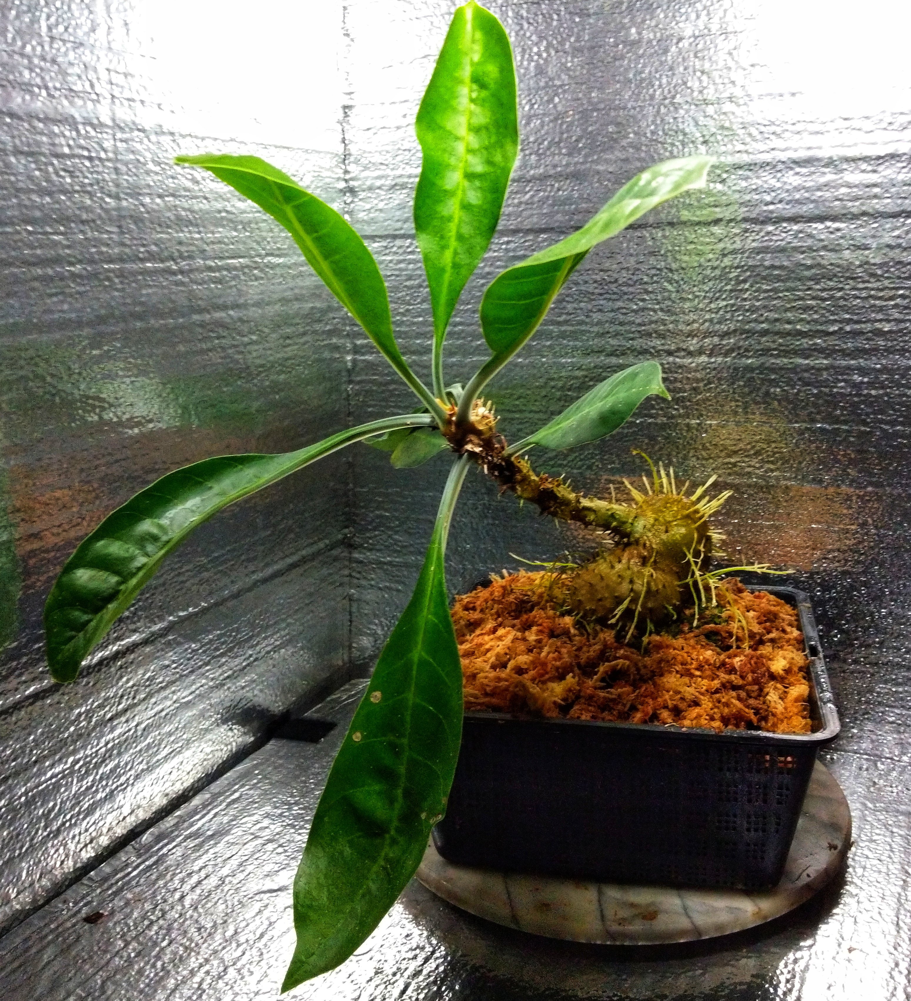
Here is the entire plant. The largest leaf is 8x23 cm with the petiole. The base spines are club shaped then gradually transition into single spines on the stem. There is an inline ridge of spines beneath the petioles. There aren't any clypeoli. Alveoli pairs. This plant has been noted to have orange fruit and not pink fruit.


-
Myrmecodia cf. pulvinata notes and images. Here are some observations on a Myrmecodia cf. pulvinata; to see how it compares to Myrmecodia tuberosa "pulvinata" and possibly find out more about the origin, identity and characteristics. The plant has been growing in a terrarium under grow lights. It is growing fast and large. When the flowers emerge, to me, it looks like molars surfacing through gums. The characteristics I find most interesting are the translucent petioles that are sharply keeled and the clubbed spines. Myrmecodia tuberosa "pulvinata" is noted to be a heterostylous species. Some plants of the population will produce brevistyle flowers and some plants will produce longistyle flowers. After examining the flower, it appears that this particular plant is producing longistylous flowers with a long style reaching to the apex of the corolla. The anthers were expired and it was difficult to determine a location. The anthers did not have any pollen when I checked the flowers, so I have not noticed if this plant has the characteristic pollen with a single vesicle (fluid filled sac). The bottom of the corolla had a ring of hair like a whisker biscuit, like those used in archery for holding the arrow. I noticed something strange about the flower; there was a "shield" of flower tissue capping the corolla with the stigma pressed against it. More detailed images can be taken when it's next flower matures. Another thing I noticed, is that ants have been putting debris around and on top of the flowers. Also, the flowers have roots surrounding them on the alveoli rim. I think the high humidity in the terrarium could be causing the aerial roots to develop. The caudex has several aerial roots as well, the stem does not have any roots. I am not sure how Anthorrhiza derived that name but doesn't Anthorrhiza translate to flower-root? Is this a trait of Anthorrhiza also? Has anyone noticed roots growing around the alveoli and flowers of other species?


-
Here is an interesting link with images of Microgramma brunei with tuber structure details. http://www.seedsplants.kimeracorporation.com/articles/solanopteris-brunei-a-rare-ant-fern-from-costa-rica.html There are some macro images of Microgramma bismarckii I have photographed and posted. This was the most fitting place to post the images. http://myrmecodia.invisionzone.com/topic/653-myrmecophytic-microgramma-spp-with-images/?tab=comments#comment-3280 I will follow up with more images of a tuber I have preserved that I plan to section; to get more details on the natural, versus ant created boundaries of the tuber chamber formation.
-
Tubers of a cultivated Microgramma bismarckii, at different stages of growth, becoming darker as they age: A budding tuber, 4.5 mm wide. There is a small cavity in developing tubers. A green tuber in mid development. A darker tuber in advanced development, 22 mm width, excluding "horns". The same tuber photographed months earlier. The entrance is oriented in a similar position on all tubers, located on the inferior side adjacent to the rhizome connection. A cross section of a tuber would be interesting to see.


-
Pollen dimensions: ~110 μm x 54 μm. 40x objective. Myrmecodia species from Irian Jaya with winged clypeoli. Images of dry vesiculate pollen with a smooth surface.


-
false scale on myrmecodia tuberosa
Philpatrick replied to Orchidman's topic in Diseases and other problems
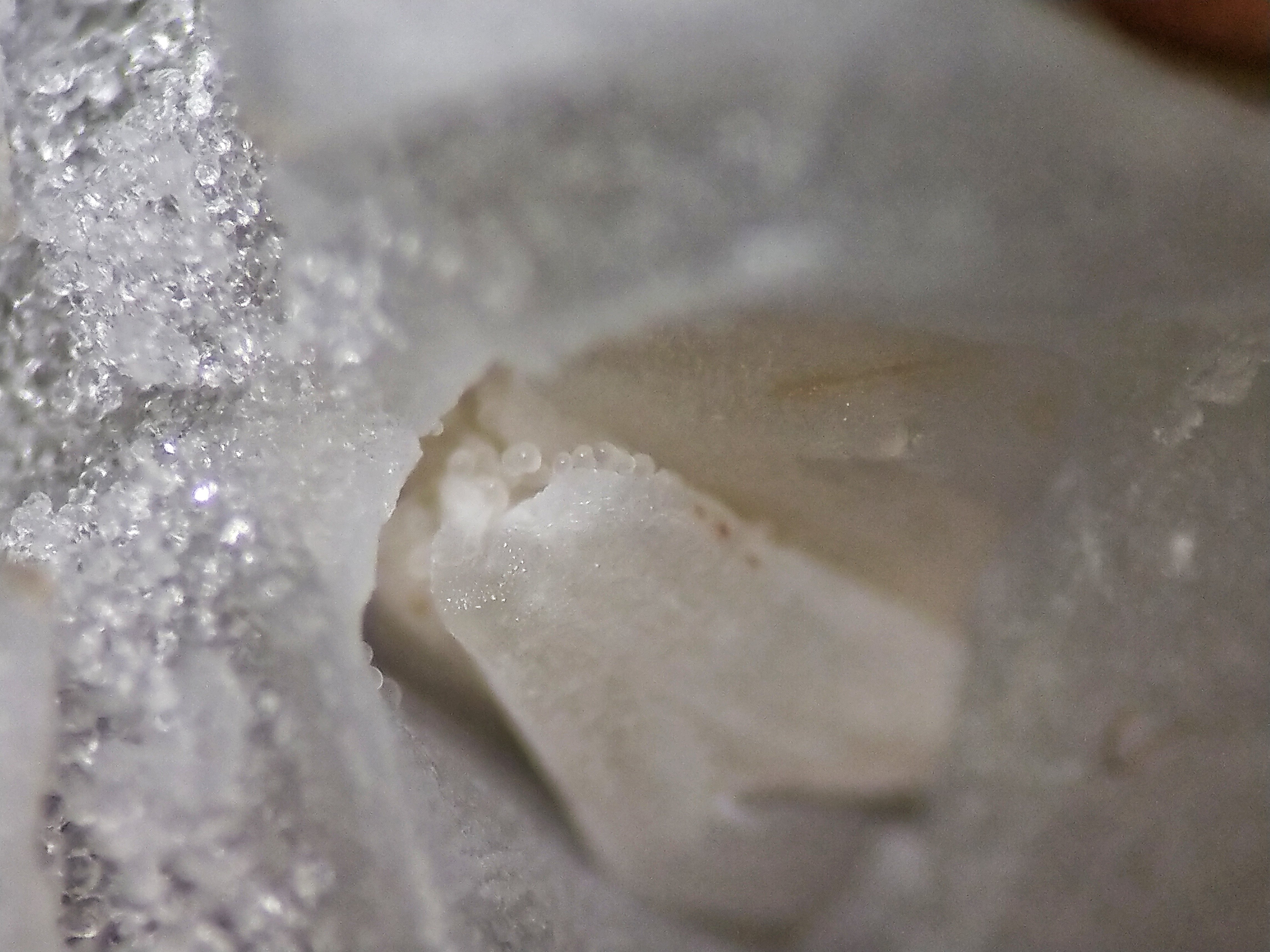
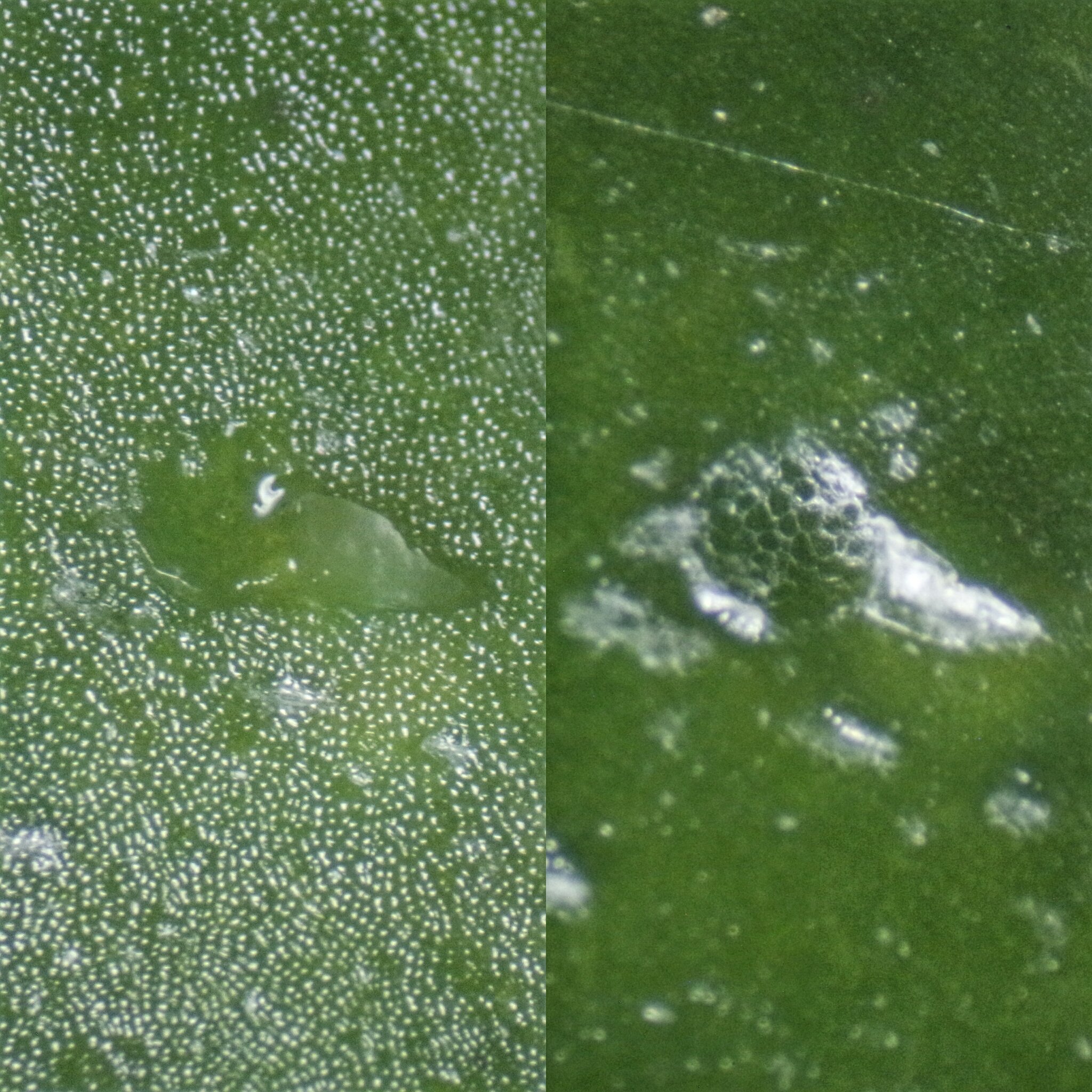
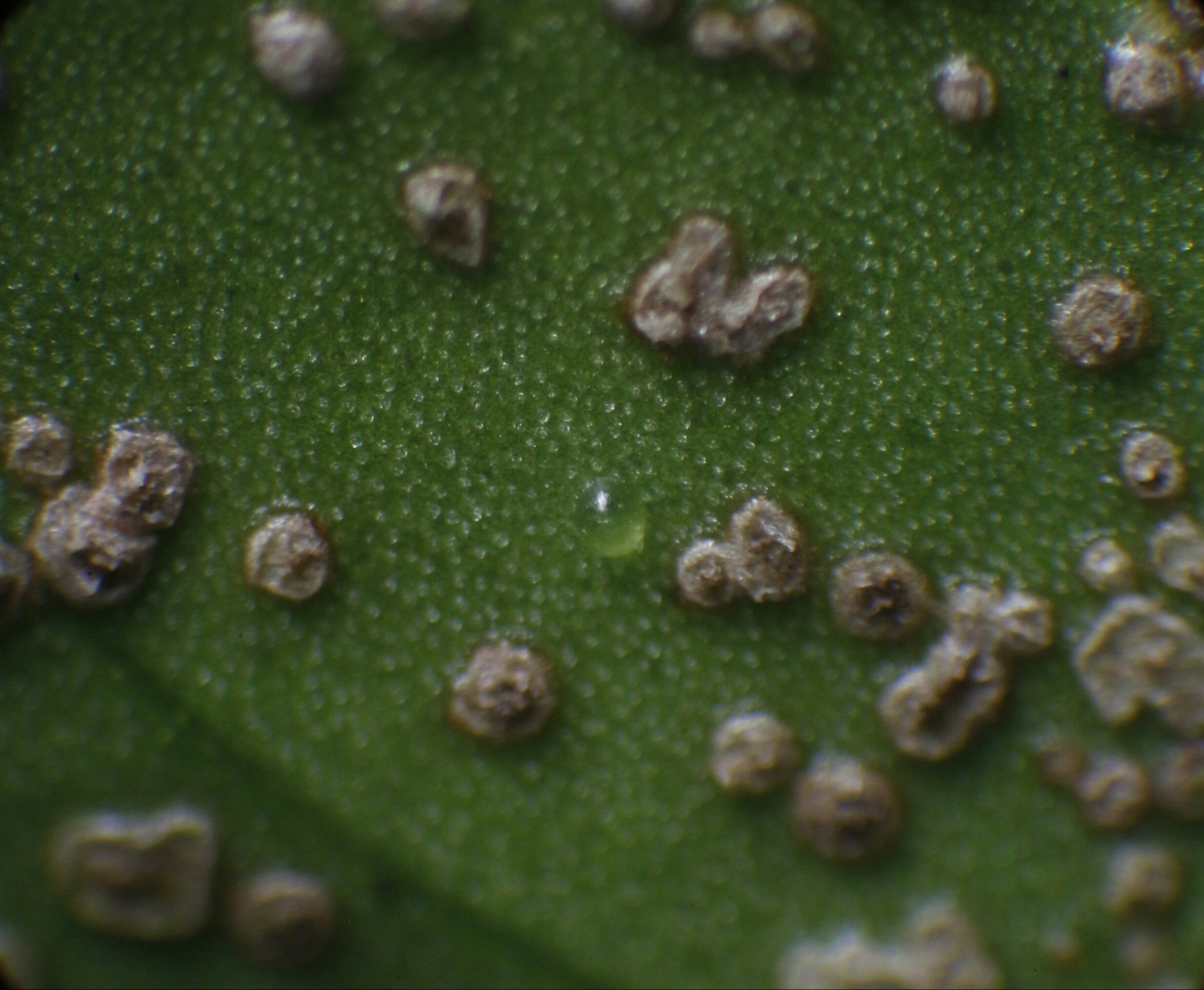
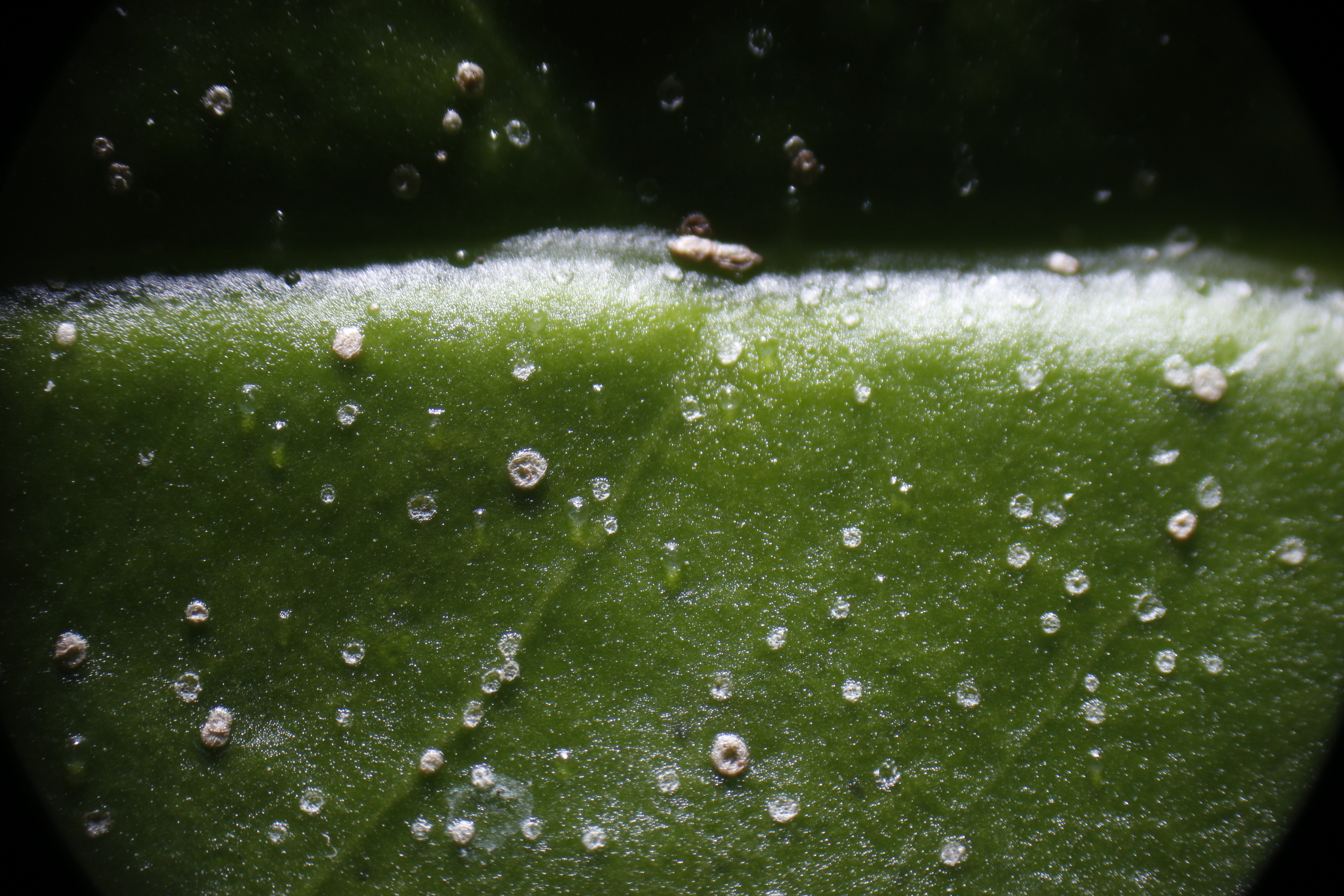
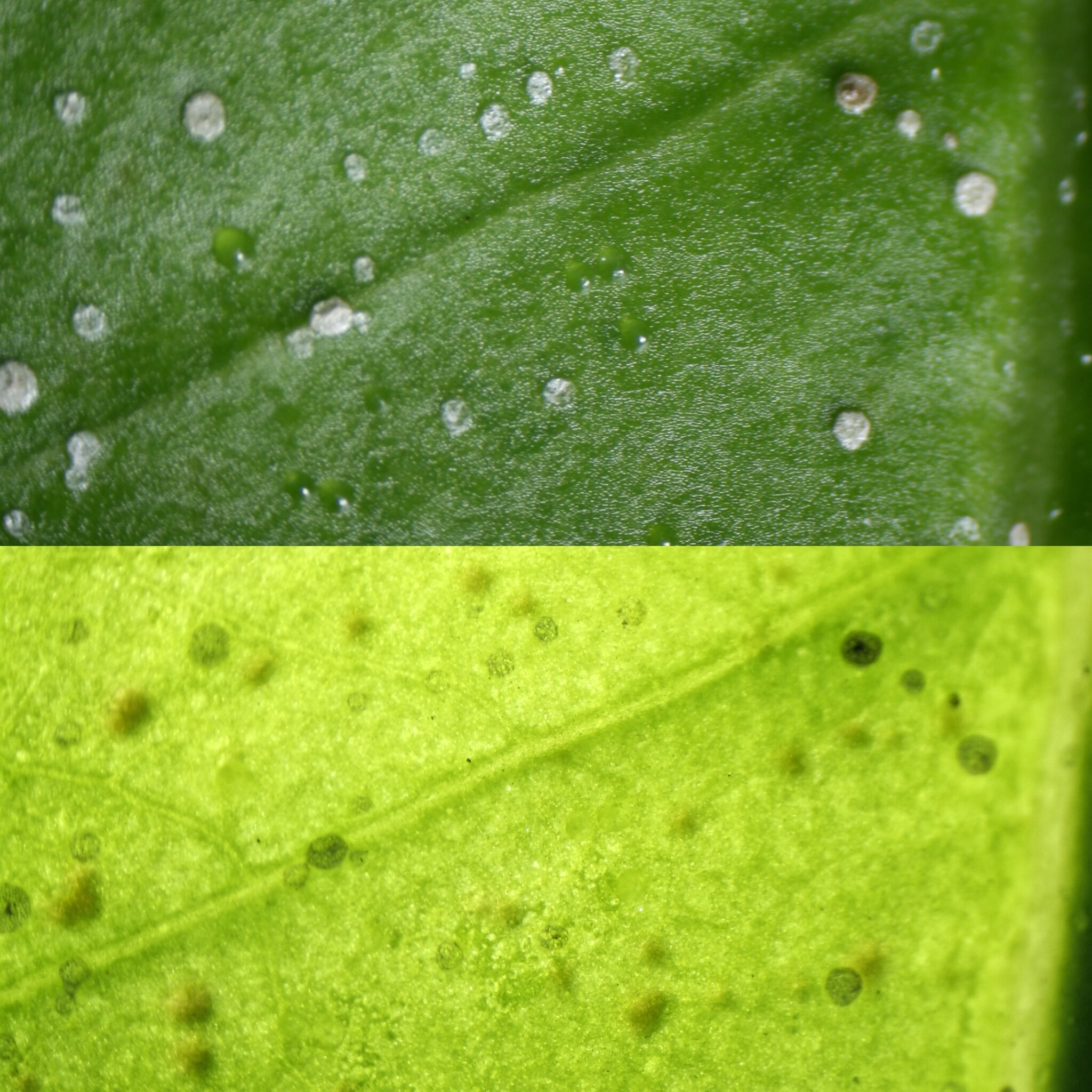
I did not record any magnification data and I will start to post that information. The microscope was a zoom stereo trinocular dissecting microscope on an articulating arm. The photoport is coupled with a camera body and a tethered flash gun is used for lighting. The articulating arm combined with a turn table makes it easy to navigate around to scope the plant. I will try to include measurements and use a microscope slide rule since the images have been resized and cropped. I took some pictures of the fluid before and after evaporation (1). I am not sure about composition. No fluid rushed out. I opened another "blister" with a hypodermic needle (2), the fluid inside quickly evaporates and large cell walls can be seen. (1) (2) When the ants explored them with their antennae It reminded me of how ants zone in on extrafloral nectaries of some Nepenthes. These are thicker areas on the undersides of the leaves (3) and (4). My guess is the thicker areas are formed when the process repeats on the same site or proximal, overlapping and creating a thicker layer. If it is cork tissue, I am working on images to visualize the suberin if present. (3) (4) Yes Frank you are correct, the top darker green photo is the flash illuminating the top of the leaf with reflected light. The lighter yellow photo is the flash transmitting light through the leaf from behind. I took these pictures as an easy way to see if the blisters were symetrical on front as well as back. The dark shadows on the lighter image are mainly cast from the growths on the backside. The blisters can only be seen as an outline. Yes, the newly formed blisters are crystal clear. The plant is in a terrarium with high humidity. I had a question Orchidman. Are the crystals on the top side of the leaf as well as the underside?


-
false scale on myrmecodia tuberosa
Philpatrick replied to Orchidman's topic in Diseases and other problems
Do these liquid filled "blisters" fit the description? I also have more images to make observations from with various types of corking. The ants that were roaming this Myrmecodia were chewing off the blisters and had to be road blocked to keep the blisters intact to photograph them. I coated the leaf petiole with petroleum jelly to barricade them. This worked, though I didn't know ants could jump to the leaf and I had to eliminate launch points. To determine if the blisters developed symmetrically on the back of the leaf, two images were taken frontlight and backlight respectively. Over time the blisters dehydrated leaving a spot with a wrinkled epidermis. More blisters formed overlapping previous blister sites.