Philpatrick
-
Posts
68 -
Joined
-
Last visited
Content Type
Profiles
Forums
Gallery
Events
Everything posted by Philpatrick
-
After reading the pages again, I found some of the spelling was different. "Cette espèce est beaucoup moins frequente que la M. Camponoti (sic) sauf de três rares exceptions, on ne Tobserve que sur les nids de Camponotusfemoratis." Suggested spelling: "Cette espèce est beaucoup moins fréquente que la M. Camponoti (sic) sauf de très rares exceptions, on ne l'observe que sur les nids de Camponotus femoratus." Suggested changes: frequente › fréquente (added accent mark "é") três › très ( changed accent mark from ê to è). Tobserve › l'observe Camponotusfemoratus › Camponotus femoratus
-
This article has some good information. https://www.researchgate.net/publication/316666507_Phylogeny_of_the_tribes_Juanulloeae_and_Solandreae_Solanaceae
-
 I really like the third image. It gives me ideas for a potluck. I'm thinking butter, sour cream, chopped green onions and bacon pieces would be good as toppings for that nice looking tater. I am glad you shared this. Amazing post!
 -
This is what two fresh pollen grains looked like under reflected light before staining and treatment.
-02.thumb.jpeg.10feb64c831da1573364b736945b7223.jpeg)
-
I had to recheck, restain and reimage a pollen sample to find that the pollen features were consistent. In the last image I think it is interesting because this pollen looks like the exine has a croton pattern, you can see the raised croton pattern especially in the last image from the set above. It looks like interconnected rings with 5/6 triangular columns on the muri. The raised areas on the exine appear darker in color. The muri is the raised area forming the reticulated pattern, the columns on the muri form the croton pattern. The lumina are the spaces between the muri. The brochus, is a lumen ( singular of lumina ) including half the width of the muri. To measure a brochus ( singular of brochi ), measure a lumen including half the width of the muri; halfway into the muri surrounding the lumen. The measurement of that space, the brochus, could be useful to know. The pollen has an exine pattern that closely resembles a croton pattern, but it could be another type of exine patterning. More resolved images will help identify this. Hydrated, stained, transmitted light brightfield.
-011.thumb.jpg.b68495df61732ac7ca5fa7eb18b9e9ba.jpg)
-
These are amazing pictures. This plant does not look like it is from this planet.
-
Widefield fluorescence micrograph of hydrated Squamellaria pollen. The pollen above was collected from a plant identified as Squamellaria. It has been stained to distinguish features. The linear aperture, the colpus (colpi plural), emits a yellow color on the pollen circumference. The red color is the exine. Dry Squamellaria pollen imaged with reflected light. In the autofluorescence image above the pollen is fresh and has not been stained. The pollen appears to be tricolpate, meaning there are three colpi. The cross section of the pollen in the polar orientation shows the three colpi simultaneously (blue arrows). The colpi are the three furrows in the circumference of the pollen wall. Stained pollen surface detail. I am working on more detailed images.
_result.thumb.jpg.c5db71c18439d2549bb8f872c3444ccb.jpg)
_result.thumb.jpg.40e45ed791ba60a3f3509c4daf87ff69.jpg)
_result-01.thumb.jpeg.e626f744a3d14b9da81638855555591c.jpeg)
_result-01.thumb.jpeg.d978237cda1192627f705a87ca93f5fa.jpeg)

-
Microgramma spores
Philpatrick replied to Philpatrick's topic in Lecanopteris, Solanopteris and other Ant associated Ferns
Microgramma bismarckii spore under reflected light._result-01.thumb.jpeg.5647b0f9c9eeff5b4ab8a15d8426d123.jpeg)
-
Echinate spore of Microgramma bismarckii under incident darkfield illumination.
_result-01.thumb.jpeg.94c6e441b90083b591c90f42d6e7df48.jpeg)
-
What type of lighting does this plant receive? What is the measured humidity in the plants growing environment? https://www.google.com/url?sa=t&source=web&rct=j&url=https://plantpathology.ca.uky.edu/files/ppfs-or-h-05.pdf&ved=2ahUKEwiXofPembDlAhW1Ln0KHWTTD0MQFjAHegQICRAB&usg=AOvVaw20KcWIT3EFgTShKlOdJJUY I have had plants with blister like growths on the leaves; not so much on the petioles like your plant is showing. A lab could test your plant to ensure it isn't a pathogen. Could it be a pathogen, the nature of the plant, or caused by adverse growing conditions? It could be oedema. You could quarantine the plant until a solid diagnosis is confirmed.
-
Myrmecodia beccarii on the Gulf of Carpentaria coast.
Philpatrick replied to Derrick's topic in Myrmecodia (Rubiaceae)
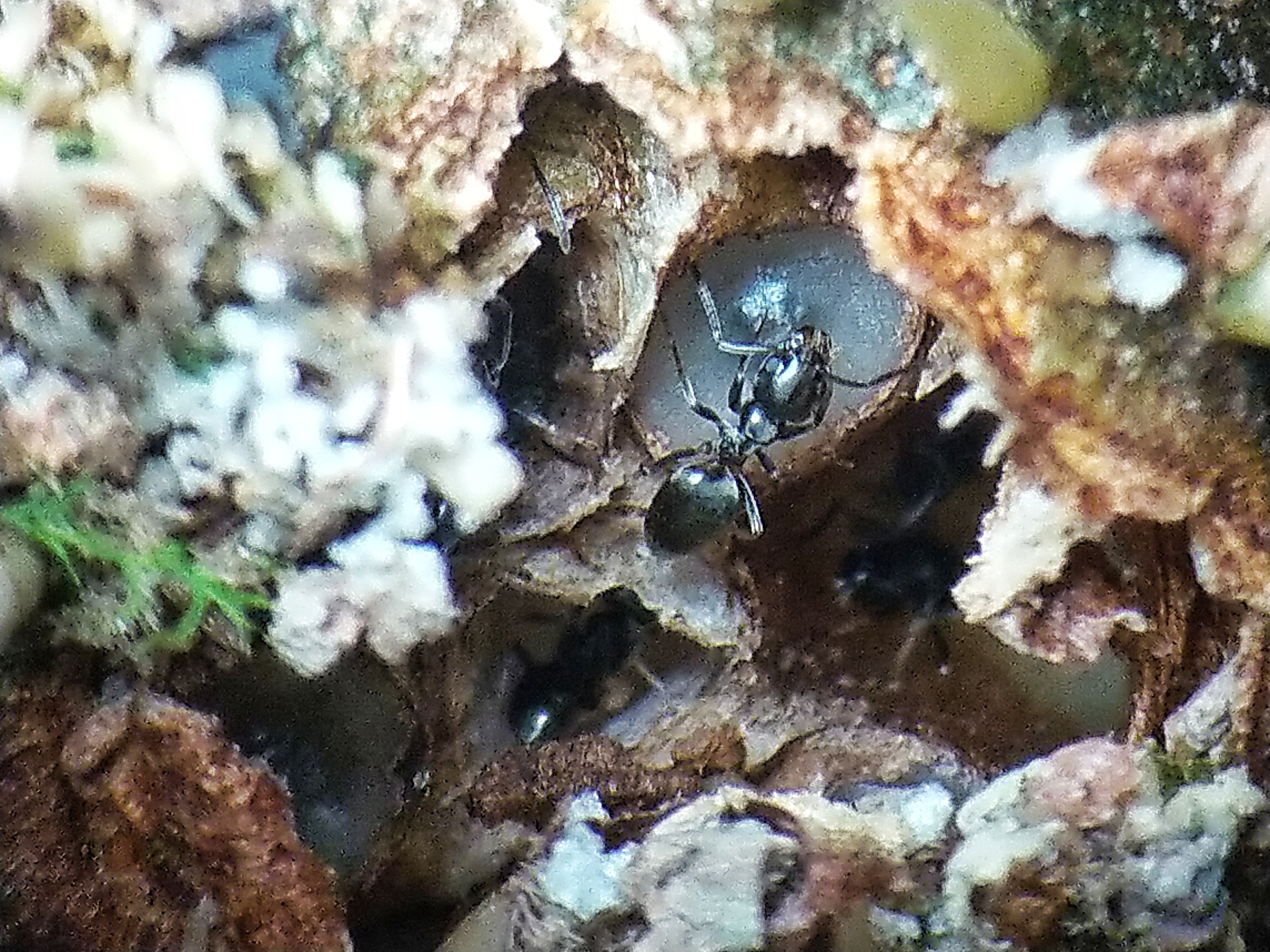
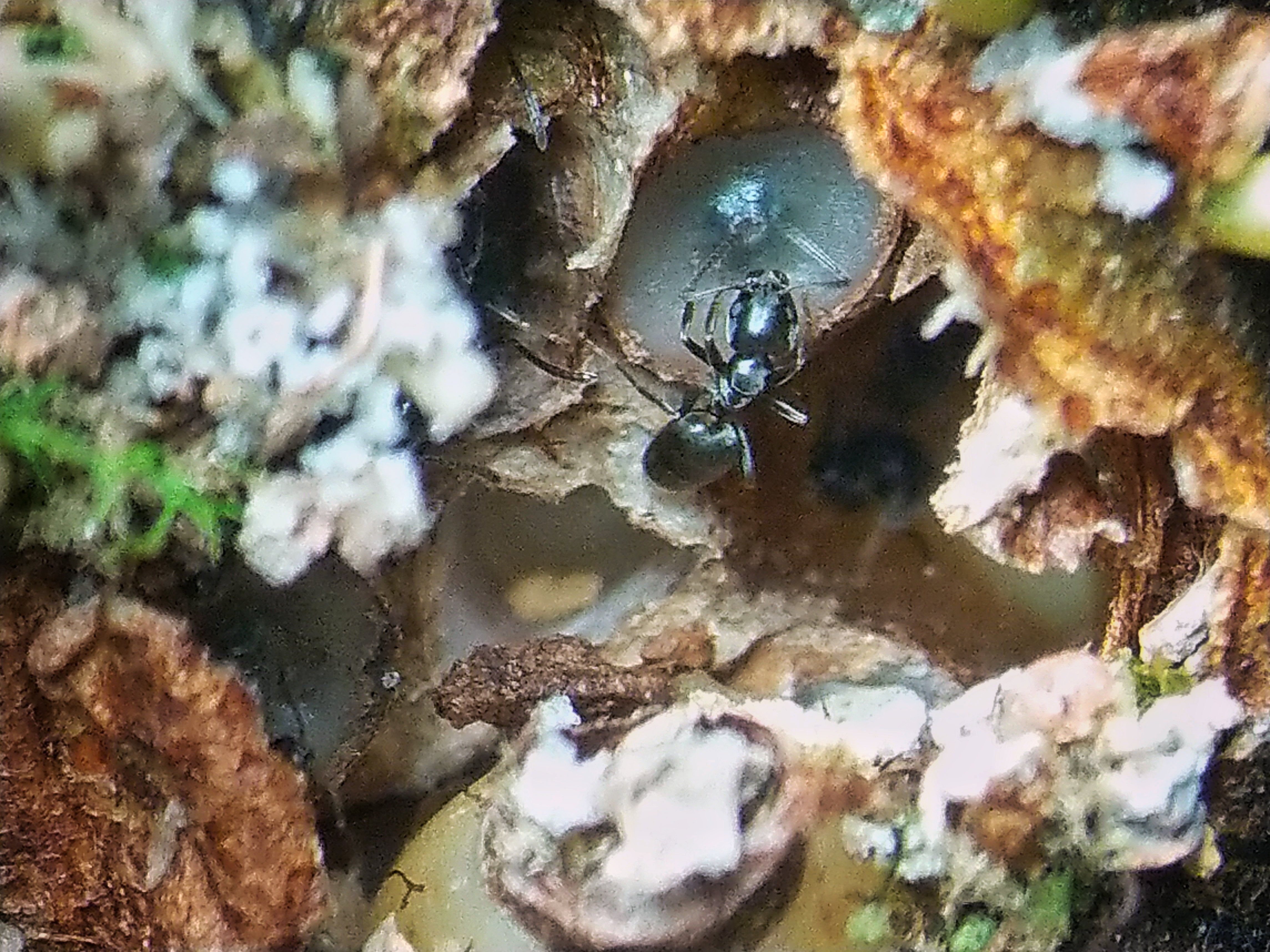
The hole is more honorable than the patch. -
According to this link, not all Squamellaria utilize the same metabolic pathway. https://journals.plos.org/plosone/article/figure?id=10.1371/journal.pone.0151317.t001
-
Myrmecodia beccarii on the Gulf of Carpentaria coast.
Philpatrick replied to Derrick's topic in Myrmecodia (Rubiaceae)
Occurrence records for Myrmecodia beccarii: https://biocache.ala.org.au/occurrences/search?taxa=Myrmecodia+beccarii&q=&fq=&wkt=&lat=&lon=&radius=&offset=20&max=20 -
Myrmecodia beccarii on the Gulf of Carpentaria coast.
Philpatrick replied to Derrick's topic in Myrmecodia (Rubiaceae)
The link above is not working: Error: unknown No record found with id: 151fcb20-a39b-4cba-b8a8-ccf03066bf33 -
The seeds from the fruit germinated and are developing nicely. The seedlings have large cavities compared to their size. At first there is not an opening and the hypocotyl is smooth. The thin surface of the caudex, where the opening is forming underneath, sheds away to reveal a cavity at their base.


-
Thank you. The drops are about a millimeter across, very noticeable by the way they refract light, and more frequent on fronds with sori. As a test, some ants were placed on a frond and they consumed the drops, with some of the meal leftover adhering to them. That plant would be very interesting to see. The informative Neotropical blueberry article has great images with amazing color.
-
According to a phylogenetic tree of plants with extrafloral nectaries (foliar nectaries for non flowering plants); there are several ferns in the polypodaceae family with foliar nectaries. I rechecked the L. curtisii again and it does have some drops forming. The location of the drops matches the location of the nectaries of several other polypod ferns. Also in another article: "Bracken nectaries are present and active on both young and mature fronds, whereas Polypodium nectaries are active only on the young fronds." (American Journal of Botany 85(5): 736–739. 1998.). This link has interesting information on fern nectaries: https://www.botany.one/2014/07/sweet-nectar-gives-ferns-bitter-taste/
-
I have had this fern for three years. There is an interesting sticky substance produced in perfect single spherical drops at almost every junction at the base of the leaflets. Detailed image and some structures dotted in a line above the drops. Some of the drops are a light sticky fluid, and some are like sticky rubber spheres. A lecanopteris curtisii is growing alongside this plant in the same conditions but I have not noticed the drops. Update: The L. curtisii has recently formed a few drops. Could this be a result of guttation, or possibly nectaries?
-01.thumb.jpeg.c65c93b59e0f1e0d12fa4989ea00b7c1.jpeg)
-01.thumb.jpeg.f011341e2ce2821d50fa3bacd499e44c.jpeg)


-
Here's one of the piles of debris the ants placed around the alveoli. They seem very content and happy. As the fruit expands outward the ants progressively remove the rim on the fruit, the developing fruit are unharmed. They explore the flat top of the fruit often with their antennae. The pile is basically every component of the complex potting media I used for this plant along with what looks like flower pieces ( It is actually ant exoskeleton molts ) and moss. There is also a pollen sac in one cavity. 12 hours later, the fruit in the lower part of the image above is already expanding and ripening.


-
The small green unripe fruit did grow and become orange quickly! Literally overnight as mentioned in this post: http://myrmecodia.invisionzone.com/gallery/image/19-m-salomonensis-ripe-fruit/
-
I had flared out the stigma lobes to get a stigma lobe count. The three lobe count was actually from another myrmecodia; this other plant has a stigma with six lobes but fused in pairs to look like three lobes. I had it stored in an unlabelled vial. I wasn't sure if it was from this plant, now I know it is not. The flower image above is from a freshly extracted Myrmecodia cf. pulvinata flower that was not stored in an unlabelled vial and no room for error. At first the stigma resides closed in the space between the ring if hairs and the anthers. Then as the anthers expire the stigma elongates and opens into the space where the pollen and anthers are and reaches to the apex of the floral chamber. Much like a protandrous flower. I have read that protandry is also a method to spatially facilitate the development of the pollen release stage and then the stigma receptivity stage; to allow each stage to have the space to develop properly.
-
It has five lobes. And an orange fruit.

-
It looks like the stigma has 3 lobes.
-
Myrmecodia pollen with two protoplasmic vesicles, one on each side (swollen areas on the left and right), and reticulate exine. Exine reticulation detail.


-
The plant I am growing looks like the plant in the second photo with the rounded leaves and is labeled the same. This plant has been stable and growing very slowly for me also. At first it lost the lower leaves. In almost one years growth It has the same pair of leaves with a new set finally emerging. It is kept in highland conditions on the dry side. Compared to the growth of the plants in the above two photos, I think my plant would greatly benefit with an environmental change as well.
-02.jpeg.5ab7b6015efdfe86a219930d79f46342.jpeg)
-011.jpg.ffa83e9e80baba8b584aa1b958df5a83.jpg)
_result.jpg.555fd81c16d311f0da6a740325336e3b.jpg)
_result.jpg.6a233d200cd0c7dab01236ffb7547e9d.jpg)
_result-01.jpeg.06b6787ba30fd86a3c457b209fba45fd.jpeg)
_result-01.jpeg.be35fa39b6f2ddd99dfe1941eb60b684.jpeg)

_result-01.jpeg.f29fce8f469297743deee668dbcb3356.jpeg)
_result-01.jpeg.7d84aa8ca647d09e6da5dc7e8027d236.jpeg)


-01.jpeg.9a4583c205da6690725116300780b5fb.jpeg)
-01.jpeg.20ee0b64fa580c501c94cd921e4ee76b.jpeg)