

Philpatrick
-
Posts
68 -
Joined
-
Last visited
Content Type
Profiles
Forums
Gallery
Events
Posts posted by Philpatrick
-
-
Tubers of a cultivated Microgramma bismarckii, at different stages of growth, becoming darker as they age:
A budding tuber, 4.5 mm wide. There is a small cavity in developing tubers. A green tuber in mid development.
 A darker tuber in advanced development, 22 mm width, excluding "horns".
 The same tuber photographed months earlier.
 The entrance is oriented in a similar position on all tubers, located on the inferior side adjacent to the rhizome connection.A cross section of a tuber would be interesting to see.
 -


Pollen dimensions:
~110 μm x 54 μm.
40x objective.
Myrmecodia species from Irian Jaya with winged clypeoli.
Images of dry vesiculate pollen with a smooth surface.
-
I did not record any magnification data and I will start to post that information. The microscope was a zoom stereo trinocular dissecting microscope on an articulating arm. The photoport is coupled with a camera body and a tethered flash gun is used for lighting. The articulating arm combined with a turn table makes it easy to navigate around to scope the plant. I will try to include measurements and use a microscope slide rule since the images have been resized and cropped.
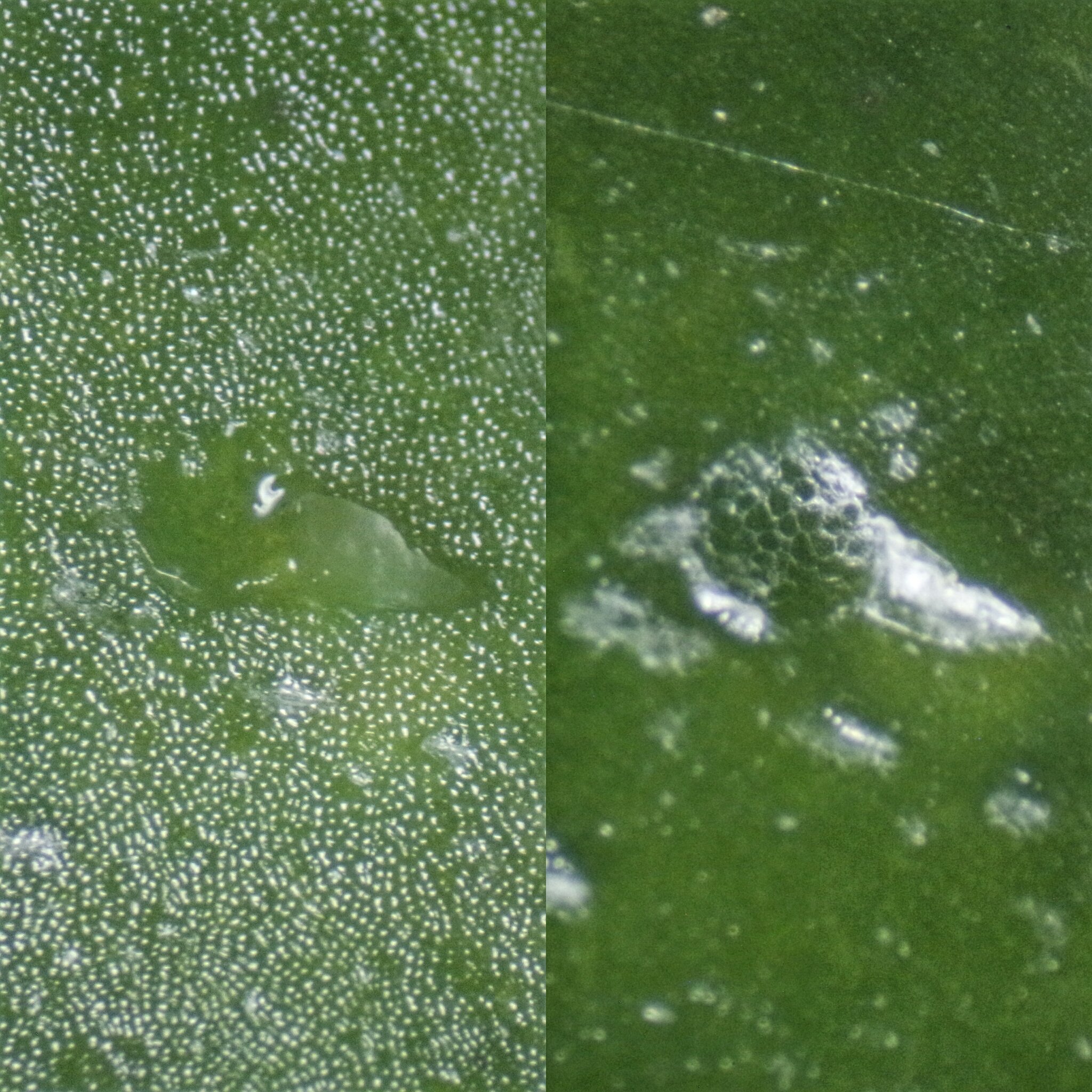
I took some pictures of the fluid before and after evaporation (1). I am not sure about composition. No fluid rushed out. I opened another "blister" with a hypodermic needle (2), the fluid inside quickly evaporates and large cell walls can be seen.
(1)

(2)

When the ants explored them with their antennae It reminded me of how ants zone in on extrafloral nectaries of some Nepenthes.
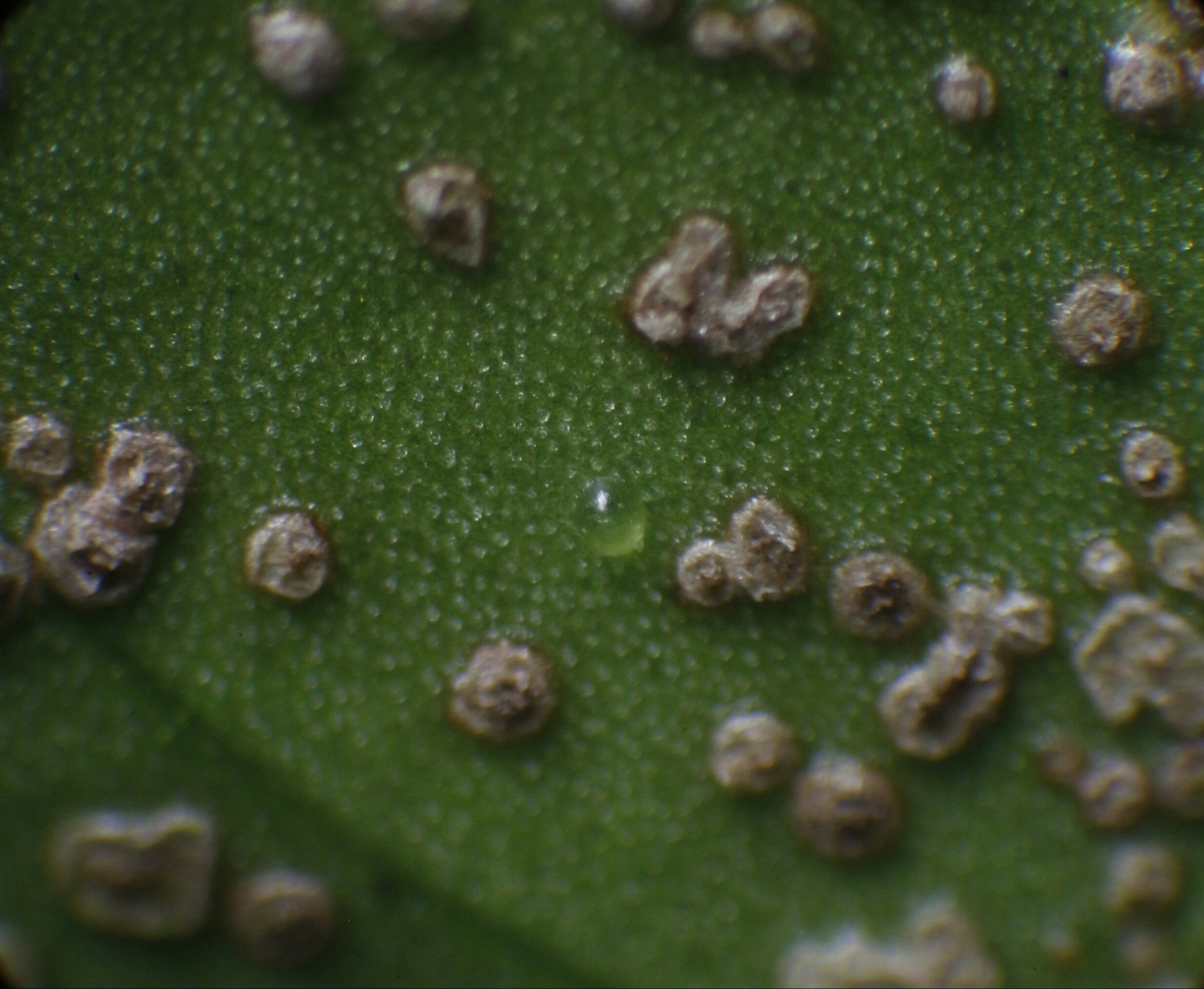
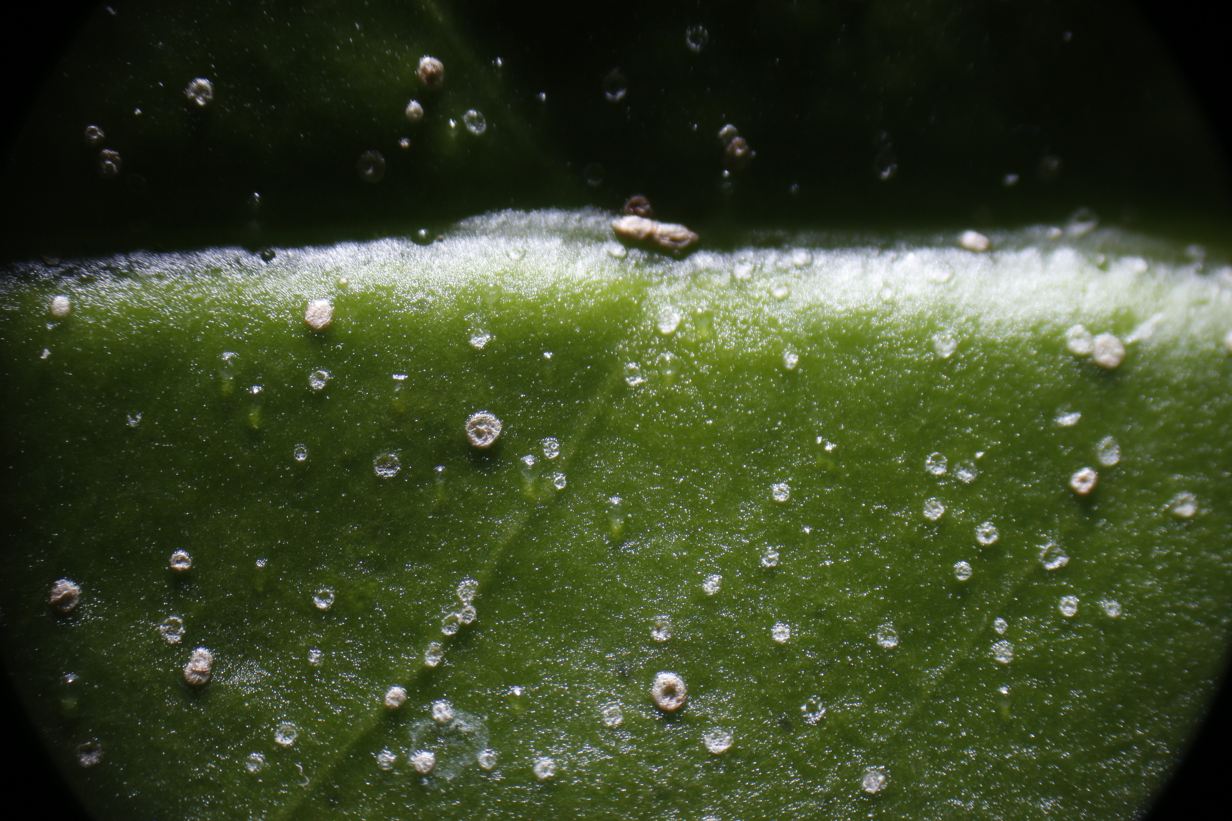
These are thicker areas on the undersides of the leaves (3) and (4). My guess is the thicker areas are formed when the process repeats on the same site or proximal, overlapping and creating a thicker layer. If it is cork tissue, I am working on images to visualize the suberin if present.
(3)

(4)

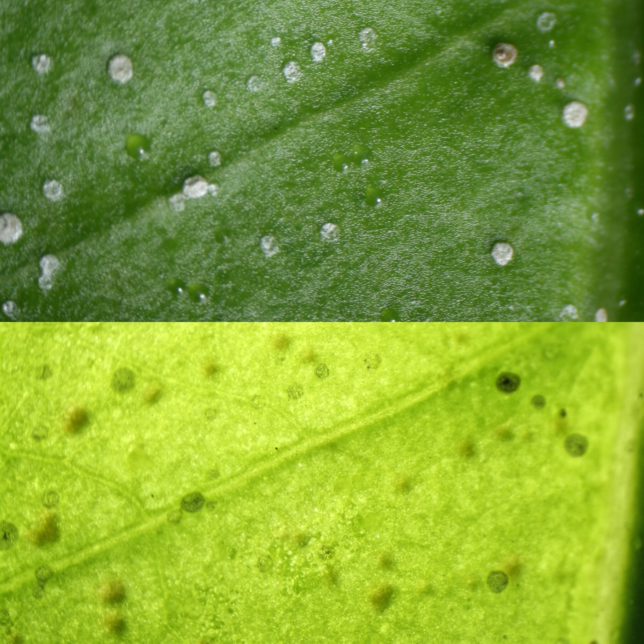
Yes Frank you are correct, the top darker green photo is the flash illuminating the top of the leaf with reflected light. The lighter yellow photo is the flash transmitting light through the leaf from behind. I took these pictures as an easy way to see if the blisters were symetrical on front as well as back. The dark shadows on the lighter image are mainly cast from the growths on the backside. The blisters can only be seen as an outline.
Yes, the newly formed blisters are crystal clear.
The plant is in a terrarium with high humidity.
I had a question Orchidman. Are the crystals on the top side of the leaf as well as the underside?
-
Do these liquid filled "blisters" fit the description? I also have more images to make observations from with various types of corking. The ants that were roaming this Myrmecodia were chewing off the blisters and had to be road blocked to keep the blisters intact to photograph them. I coated the leaf petiole with petroleum jelly to barricade them. This worked, though I didn't know ants could jump to the leaf and I had to eliminate launch points.
To determine if the blisters developed symmetrically on the back of the leaf, two images were taken frontlight and backlight respectively.
Over time the blisters dehydrated leaving a spot with a wrinkled epidermis. More blisters formed overlapping previous blister sites.


Solanopteris brunei - from Colombia - Info:
in Lecanopteris, Solanopteris and other Ant associated Ferns
Posted
Here is an interesting link with images of Microgramma brunei with tuber structure details.
http://www.seedsplants.kimeracorporation.com/articles/solanopteris-brunei-a-rare-ant-fern-from-costa-rica.html
There are some macro images of Microgramma bismarckii I have photographed and posted. This was the most fitting place to post the images.
http://myrmecodia.invisionzone.com/topic/653-myrmecophytic-microgramma-spp-with-images/?tab=comments#comment-3280
I will follow up with more images of a tuber I have preserved that I plan to section; to get more details on the natural, versus ant created boundaries of the tuber chamber formation.